
Tags
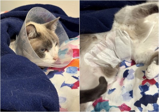
This just in… Sonia has sent me the results from Luna’s biopsy. The two tumours were malignant but Stage 1, and as they were completely resected there is a low risk of metastasis. No further treatment required. I was going to take her in to get her bandage changed this morning but it was bucketing down. Also during the night the bandage mostly came off so I found these stick-on gauze ones in the cupboard which seem to be doing the trick, and Luna is more comfortable not having a full-torso wrap-around one. The two wounds were dry and clean, and she is still taking antibiotics, so Sonia and I decided to just leave her until next Tuesday when she will have the stitches removed. Unfortunately she still needs to wear the dread cone but she seems to be taking it in stride. So, not a bad Christmas gift for my old girl.
HISTOPATHOLOGY: In the two mastectomy specimens submitted, two nodular neoplastic proliferations of the mammary gland of simple/complex nature were observed. The neoplastic epithelial cells in these nodules are arranged in acinotubular structures with sometimes irregular lumens, lined by two to four layers of cuboidal or columnar cells, with rounded, hyperchromatic nuclei and occasionally showing one or two prominent nucleoli. The degree of cellular atypia and mitotic count are low (4-5 mitoses/10 high-power fields). In one of the nodules, associated with the epithelial component, a proliferation of myoepithelial cells in solid nests with typical characteristics is observed. Both tumours show expansive growth and have been completely resected. No evidence of vascular tumor embolization is seen in the histological sections of the submitted specimens.
DIAGNOSIS: Simple/complex tubular adenocarcinomas of the breast, grade I (x2).
PROGNOSIS: Less serious.
COMMENTS: These are two breast tumours of simple/complex nature with histological characteristics that generally confer a low grade of malignancy (grade I). The absence of tumour vascular emboli in the submitted specimens indicates a low probability of distant metastasis from these tumours (regional lymph node biopsy is always recommended to confirm this assessment). Both tumours were completely resected. Finally, because mammary tumours in cats can occur in multiples, periodic monitoring of the animal is recommended for the possibility of new mammary tumours appearing.

That path report is making my eyes cross, and I’m a dedicated reader of lab reports. I’m just glad the bottom line is no worse than it is. Poor girl, having to put up with all that. I want to give her a head skritch.
LikeLike
She’s certainly been a GOOD GIRL this past week. I may let her take occasional breaks from the cone. Just five more sleeps until it comes off.
LikeLike